Histology platform
- Automated slide-scanning.
- General histology as a service: cryosectioning, vibratome sectioning, mounting, IHC, RNAscope, H&E, etc.
- Optimising new antibodies and protocols. Antigen retrieval.
- Teaching/training of histological techniques.
- Advice on tissue clearing and lightsheet microscopy.
- Training and use of our Bright Cryostat and Leica VT1200 vibratome.
Equipment
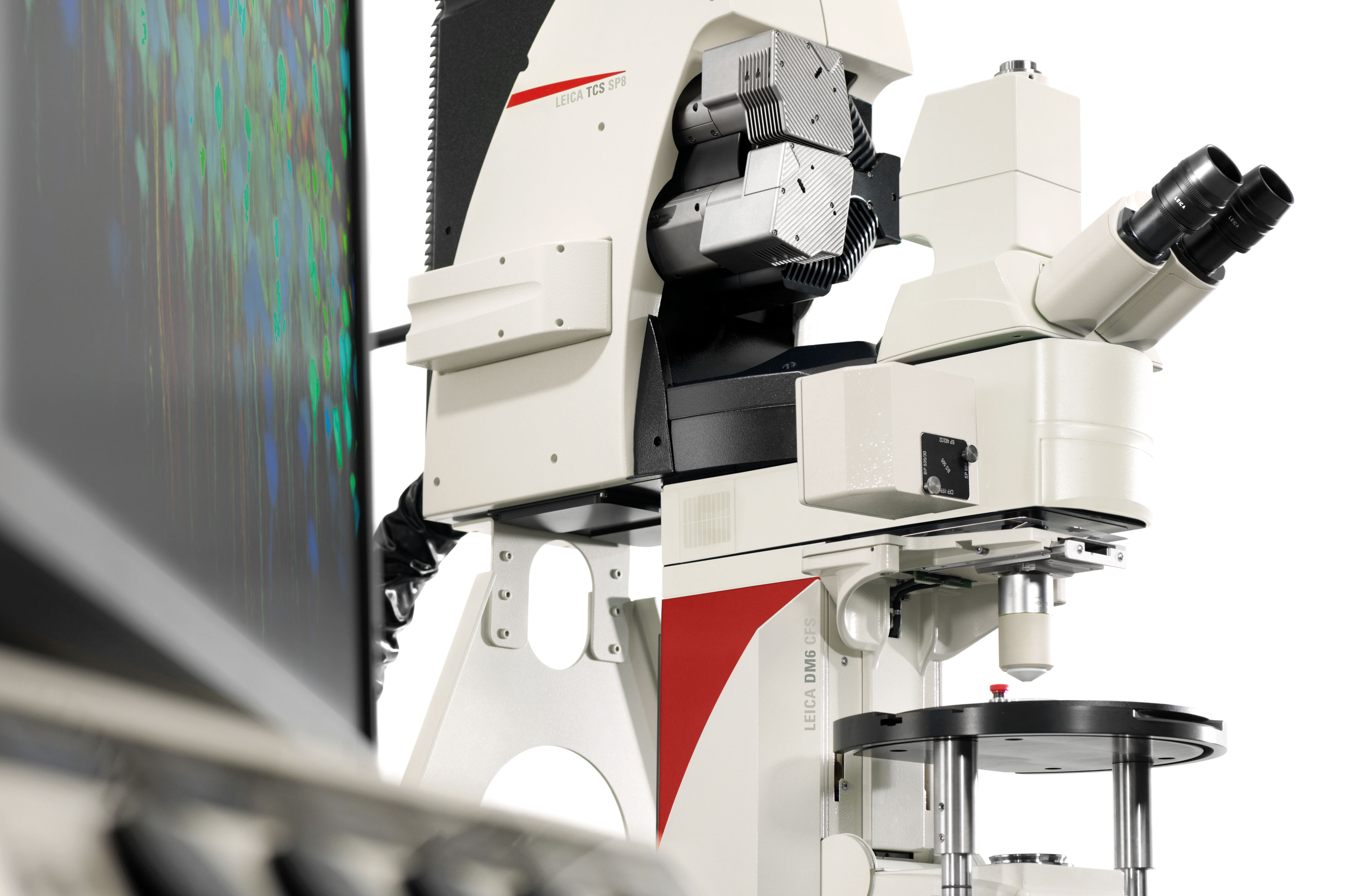
Leica SP8 Confocal
The thin optical sections produced by this confocal are ideal for imaging sub-cellular structures, such as axons and synapses. The microscope is also used for generating detailed images of cells, allowing their processes to be traced. The software has a slide-scanning feature, which allows even thick cleared slices to be imaged with ease. A white light laser with tunable excitation filter and three detectors (2x HyD and 1x PMT) with tunable emission filters provide enormous flexibility.
AMF SP8 Details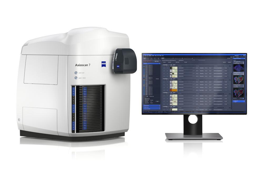
Axio Scan slide scanner
This high-throughput widefield microscope accommodates up to 100 slides, which it images automatically after an interactive setup process. The system handles both fluorescence and conventional brightfield. We run this microscope alongside our histology service, but it can also be booked and used independently.
AMF Axioscan Details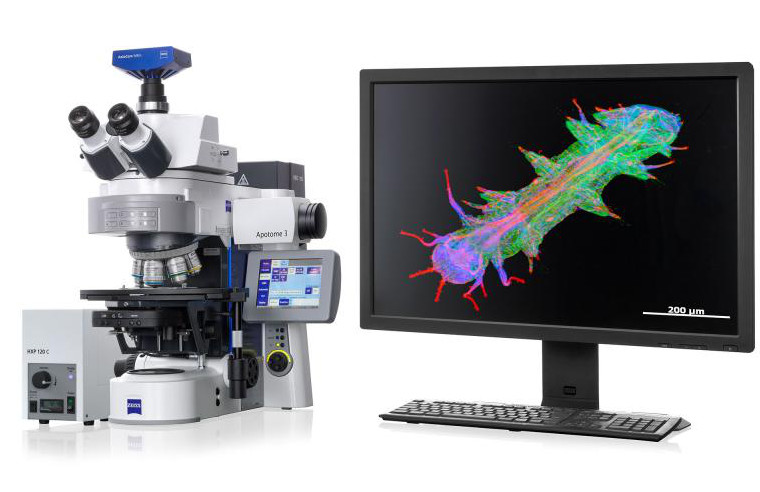
Widefield microscopes
We have two Zeiss Imagers, equipped with Apotomes for optical sectioning. These microscopes are ideal for imaging small numbers of slides interactively and have tile scanning ability. Please contact the AMF for an induction if you are unfamiliar with them.
AMF Zeiss Imager Details