MesoSPIM v5
System details and operating notes for the AMF lightsheet
Overview
We have a mesoSPIM v5 lightsheet designed by Fabian Voigt in the Helmchen Lab, Zurich. The system is based around an Olympus zoom macroscope and is ideal for screening large numbers of samples and visualising sparse cell populations, plaques, or blood vessels. The system is unsuited for samples where resolutions of about a micron or better are needed. Images are acquired with a Hamamatsu ORCA Flash 4.0. Two iChrome MLE laser launches provide excitation wavelengths of 488 nm, 561 nm, 640 nm, and 685 nm. Emitted light is filtered using a 10 position Ludl filter wheel. Open source Python code runs the microscope.
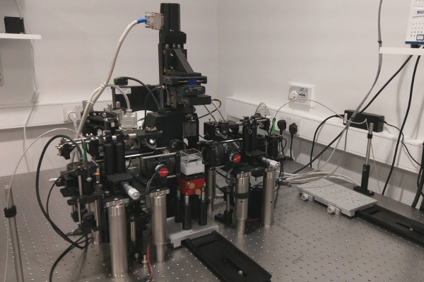
Users must be trained by the AMF before they can access the microscope, and are responsible for clearing their own samples. We can provide protocols and advice — see the clearing techniques below.
Basic usage
Turning on the system
- Turn on the Hamamatsu camera with the switch at the back of it.
- Turn the keys of each laser launch clockwise by 90 degrees. Each laser will play a little tune once it has started up.
- Start the mesoSPIM software via the icon on the Desktop.
Turning off the system
- Turn off the camera.
- Switch off the lasers with the key switch.
- Close the software.
- Cap the large cuvettes so dust does not get in.
System details
Filters
| Pos. | Use | Filter ID |
|---|---|---|
| 1 | Quad line | ZET405/488/561/640mv2 |
| 2 | GFP | FF01-520/35 |
| 3 | tdTomato & mCherry | BLP01-594R |
| 4 | — | BLP01-633R |
| 5 | 700 nm low pass | — |
| 6–9 | Unassigned | |
| 10 | Open port | n/a |
The following filters are available but not installed in the wheel:
|
|
Zooms
The mesoSPIM uses an Olympus macroscope zoom body. A stepper motor turns the zoom knob via the software. The zooms correspond to the following fields of view and pixel sizes.
| Zoom | FoV (mm) | Pixel size (mm) |
|---|---|---|
| 0.63 | 21.56 | 0.0105 |
| 0.80 | 16.86 | 0.0082 |
| 1.00 | 13.42 | 0.0066 |
| 1.25 | 10.79 | 0.0053 |
| 1.60 | 8.36 | 0.0041 |
| 2.00 | 6.69 | 0.0033 |
| 2.50 | 5.34 | 0.0026 |
| 3.20 | 4.16 | 0.0020 |
| 4.00 | 3.29 | 0.0016 |
| 5.00 | 2.62 | 0.0013 |
| 6.30 | 2.12 | 0.0010 |
The NA varies with zoom: NA = 0.075 at 1× zoom and NA = 0.22 at 4× zoom. The change in NA keeps pace with magnification so the image does not dim up to about zoom 4. Beyond zoom 4 the NA no longer increases, so the image dims. NA 0.22 corresponds to a lateral resolution of about 1.4 microns.
Useful links
Reviews
- Microscopy: seeing through tissue by Vivien Marx (2014)
- Clarifying tissue clearing by Richardson & Lichtman (2016)
- Physical and chemical mechanisms of tissue optical clearing (2021)
- Tissue clearing and its applications in neuroscience (2020)